

₨3,200
Package includes: Set of 3 pieces
Root Tip Forceps are specialised extraction instruments designed exclusively for grasping, manipulating, and retrieving broken root tips, retained root fragments, and residual roots from alveolar sockets. Oral surgeons and general dentists encounter root fracture as one of the most common complications during tooth extraction — and when standard forceps and elevators cannot deliver the fragment safely, the Root Tip Forceps provides the fine, narrow beak geometry needed to engage and retrieve root remnants from the confined apical portion of the extraction socket. Because leaving retained root fragments generates post-surgical infection risk, delayed healing, and patient complaints, having the correct root tip forceps immediately available transforms a potentially difficult complication into a manageable, predictable clinical step.
In addition to root fracture management, dental root tip forceps serve planned procedures — removing intentionally retained roots in geriatric patients, retrieving deciduous root fragments in paediatric patients, and extracting deeply submerged root tips following crown separation. As a result, these instruments belong on every oral surgical extraction tray as a standard component rather than a specialty item reserved only for emergencies.
A dental root tip forceps is an extraction forceps with extremely fine, narrow, pointed beaks designed to enter deep alveolar sockets and engage small root fragments that conventional extraction forceps beaks — designed for intact crown and root anatomy — cannot grip. The defining design characteristic of root tip pick forceps is the beak profile — narrow enough to access the apical third of an extraction socket alongside bone walls, yet strong enough to apply controlled gripping and rotational force to a root fragment without beak deformation or fracture.
Unlike standard extraction forceps that close around the full root trunk, the root tip forceps closes around a root fragment whose coronal surface lies at or below the alveolar crest — a position that places it entirely beyond the reach of conventional beak designs. Therefore, the pointed fine tips of this instrument bridge the capability gap between elevators that luxate root fragments and forceps that deliver the luxated fragment from the socket after adequate mobility is achieved.
Root fracture during extraction occurs most frequently in teeth with hypercementosis, curved roots, heavily restored crowns, dilacerated apices, and in patients with dense cortical bone or brittle root structure from endodontic treatment. Moreover, any extraction performed without adequate pre-elevation with Coupland or Warwick James elevators carries elevated root fracture risk. Consequently, assembling an extraction tray that includes root tip forceps alongside elevators and standard forceps prepares the clinical team for the full range of extraction outcomes — planned and unplanned — without requiring a mid-procedure instrument search.
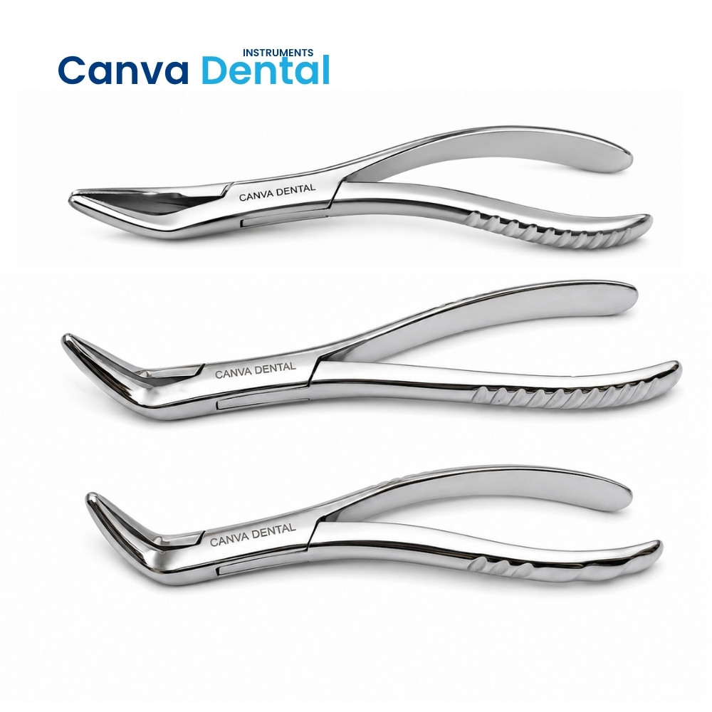
Each root tip forceps in our range delivers the beak precision, handle strength, and jaw design that retained root retrieval requires consistently across all socket depths and fragment sizes:
Several root tip forceps types exist, each designed for specific socket positions, root fragment depths, and access angle requirements. Understanding the available types helps dental teams build a complete root tip instrument set that covers every retained root scenario:
| Type | Beak Design | Best Application |
|---|---|---|
| Straight Root Tip Forceps | Straight aligned beaks | Anterior socket root tips — maxillary and mandibular incisors and canines |
| Bayonet Root Tip Forceps | S-shaped bayonet offset | Posterior maxillary root tips — reaches over the palate without handle obstruction |
| Angled / Curved Root Tip Forceps | Angled beak offset | Mandibular posterior roots — access to lower molar root fragments from correct angle |
| Universal Fine Beak Forceps | Narrow straight with slight curve | General-purpose anterior and premolar root tip retrieval |
| Beak Category | Tip Width | Root Fragment Size |
|---|---|---|
| Extra Fine | 1.5 – 2.0 mm | Very small apical fragments — curved root tips, fine anterior roots |
| Fine Standard | 2.0 – 2.5 mm | Standard anterior and premolar root fragments |
| Medium | 2.5 – 3.5 mm | Premolar and molar root fragments with larger cross-section |
Therefore, stocking a complete root tip forceps set — straight, bayonet, and angled in fine and medium beak widths — ensures the surgeon always selects the instrument that matches both the socket position and the root fragment size, rather than forcing an inappropriately wide beak into a narrow socket and risking buccal plate fracture during the retrieval attempt.
The complete range of root tip forceps uses extends across extraction complications, planned surgical procedures, and paediatric dentistry. Although retained root fragment retrieval after crown fracture is the most frequent application, root tip forceps uses cover every scenario where a confined, deeply positioned root structure requires precise forceps delivery:
The decision to leave a retained root fragment — sometimes called intentional root retention — requires careful clinical judgment supported by specific radiographic and patient criteria. However, the majority of root fracture cases during routine extraction do not meet these criteria and require immediate retrieval with retained root extraction forceps instruments.
Retained root fragments left in the alveolus after extraction create several well-documented clinical risks. Because root fragments retain viable periodontal ligament fibres and cementum, they can remain quiescent initially — only to develop periapical pathology weeks or months after extraction when the residual PDL undergoes necrotic breakdown. Moreover, retained roots in implant sites prevent bone-to-implant contact in the fragment zone, creating implant osseointegration failure that only becomes apparent several months after implant placement.
In addition, retained root fragments near the inferior alveolar canal or maxillary sinus floor carry specific anatomical risks — progressive infection in these areas reaches adjacent vital structures and generates complications significantly more complex than the original root retrieval would have required. Therefore, attempting root tip retrieval with the correctly selected root tip forceps at the time of initial extraction is almost always preferable to managing delayed complications from retained roots.
Intentional root retention — leaving a small, completely asymptomatic root fragment in situ — is clinically acceptable only when the fragment is small (less than 3–4mm), completely enclosed in bone with no periapical pathology, located away from vital structures, the patient is medically compromised with elevated surgical risk, and informed consent is documented. Furthermore, the decision requires radiographic confirmation and specific follow-up scheduling — it is not a default response to a difficult retrieval but a considered clinical decision with defined criteria.
Several instruments address retained root retrieval in oral surgical practice. Understanding how root tip pick forceps compares to alternative retrieval instruments helps clinicians build a logically organised root retrieval tray:
| Instrument | Mechanism | Best For | Limitation vs Root Tip Forceps |
|---|---|---|---|
| Root Tip Forceps | Fine beak gripping and delivery | Mobile root fragments — gripping and removing from socket | — |
| Apexo Elevator | Thin curved blade luxation | Initial loosening of root tip before forceps delivery | Luxates only — cannot grip and remove fragment |
| Warwick James Elevator | Curved blade PDL separation | Curved root luxation before root tip forceps application | Too wide for apical socket access in most cases |
| Cryer Elevator | Triangular tip inter-radicular | Post-sectioning molar root elevation | Inter-radicular use — not apical root tip access |
| Root Tip Pick | Fine pointed probe | Locating and positioning root tip before forceps | Cannot grip — probe function only |
PiezosurgeryUltrasonic vibrationBone removal around deeply impacted root tips near nervesEquipment cost — not standard tray instrumentConsequently, the most effective root tip retrieval workflow uses the Apexo elevator for initial loosening, the Root Tip Forceps for gripping and delivering the mobile fragment, and the Lucas Bone Curette for subsequent socket debridement — each instrument addressing a distinct phase of the root retrieval and socket management sequence.
Before applying the root tip forceps, confirm that the root fragment has adequate mobility — attempting forceps delivery of a firmly attached root tip without prior elevation drives the fragment deeper into the socket rather than retrieving it. Use the Apexo elevator or a fine Warwick James curved elevator to loosen the fragment through gentle rotational force before introducing the forceps beaks. Moreover, ensuring the surgical field is clear of blood through Frazier suction tip aspiration before beak insertion allows accurate visual placement of the fine beak tips at the correct position on the root fragment surface.
Insert the root tip forceps beaks into the socket alongside the bone walls — never forcing the beaks between the root and bone under excessive pressure. Position the beaks so they contact the root fragment as far apically as accessible, with the serrated inner surfaces engaging the smooth root surface on opposing sides simultaneously. In addition, close the beaks firmly before applying any rotational or tractional force — loose beak closure allows the fragment to slip from the jaws and potentially displace further apically under the retrieval force.
Apply a combination of gentle rotation and traction once the beaks achieve secure fragment contact — rotating the fragment in the direction of its natural root curvature to follow the socket path, then delivering it occlusally in the extraction direction. Furthermore, if resistance is encountered during delivery, release the forceps and return to elevator loosening before attempting a second delivery — forcing the root tip forceps against a firmly anchored fragment creates bone plate fracture risk rather than safe root retrieval.
All stainless steel Root Tip Forceps in our range withstand repeated autoclave cycles at 134°C without beak deformation, joint corrosion, or serration damage. However, the fine beak tips are the most vulnerable component — any bending, chipping, or misalignment of the beak tips reduces the instrument’s ability to access deep sockets and engage small root fragments accurately. Therefore, inspecting the beak alignment by closing the forceps under direct light and confirming both tips meet at exactly the same point before each procedure identifies tip damage before it compromises retrieval performance.
In addition, ultrasonic cleaning before autoclaving removes bone chips, PDL tissue, and blood from the beak serrations and box joint — areas that manual cleaning cannot reliably reach. Furthermore, the box joint requires periodic inspection for looseness — a loose joint allows the beaks to misregister under gripping force, causing the root fragment to slip laterally from the beak tips rather than being held securely during the delivery stroke. Consequently, replacing root tip forceps when beak misalignment or joint looseness becomes apparent protects both retrieval success and patient safety during every extraction procedure.
We supply Root Tip Forceps — in straight, bayonet, and angled beak designs across extra fine, fine, and medium beak widths for upper and lower arch root retrieval — to oral surgery departments, general dental clinics, teaching hospitals, dental colleges, and instrument distributors across Lahore, Karachi, Islamabad, Multan, Peshawar, Faisalabad, Rawalpindi, and all major cities in Pakistan. Moreover, our institutional supply team handles bulk procurement for dental college oral surgery departments and hospital surgical units at competitive pricing.
Contact our team for current root tip forceps pricing in Pakistan, complete set availability, and delivery timelines for your clinic or institution.
Q: What are Root Tip Forceps used for in dentistry?
Root Tip Forceps retrieve broken root tips, retained root fragments, and deeply positioned residual roots from alveolar sockets during and after tooth extraction. Primary root tip forceps uses include fractured apex retrieval after crown separation, hypercementosed and dilacerated root delivery, individual root retrieval after molar sectioning, deciduous root fragment removal in paediatric patients, and pre-implant site root clearance. In addition, these instruments manage planned extraction scenarios where roots require individual delivery using fine beaks rather than conventional extraction forceps that cannot access deep socket positions.
Q: How are Root Tip Forceps different from standard extraction forceps?
Standard extraction forceps carry wide beaks designed to engage the full root trunk below an intact crown — they close around the cervical root surface and deliver the tooth through buccal-lingual expansion and traction. Root tip forceps, however, carry extremely fine, narrow beaks — typically 1.5–3mm wide — designed to enter the deep apical portion of an extraction socket alongside bone walls and grip a small root fragment that standard beaks cannot access. Therefore, root tip forceps address extraction complications and anatomically challenging root retrievals that fall entirely outside the capability of conventional forceps beak geometry.
Q: What is the difference between straight, bayonet, and angled Root Tip Forceps?
Straight root tip forceps carry direct beak alignment suited for anterior socket root fragments accessible from a straight approach. Bayonet root tip forceps carry an S-shaped offset that positions the beaks over the maxillary posterior arch without the handle obstructing access — making them the standard choice for upper molar and upper premolar root tip retrieval. Angled root tip forceps carry a lateral beak offset suited for mandibular posterior root fragments where the access angle differs from the anterior straight approach. Consequently, stocking all three designs provides complete arch and quadrant coverage for every retained root retrieval scenario encountered in clinical practice.
Q: Should a root tip always be removed after it breaks during extraction?
In the majority of cases, yes — retained root fragments generate periapical pathology risk, delayed healing, implant site contamination, and patient discomfort that make immediate retrieval preferable to leaving the fragment. However, intentional root retention is clinically acceptable only when the fragment is very small, completely enclosed in healthy bone, shows no radiographic periapical pathology, sits away from vital structures, and the patient carries elevated surgical risk. Furthermore, any decision to leave a root tip requires documented radiographic assessment, patient informed consent, and scheduled follow-up radiography — it is never an acceptable default response to a difficult retrieval.
Q: Are Root Tip Forceps autoclavable?
Yes. All stainless steel Root Tip Forceps in our range withstand autoclave sterilization at 134°C.
Reviews
There are no reviews yet.